Heel pain causes
Plantar fasciitis vs heel spurs: why the spur on your X-ray is probably not the problem
An X-ray shows a bony spike on the underside of the heel, the report names it, and the conclusion writes itself: that spike is what hurts, and it needs to come out. It is one of the most persistent misunderstandings in foot and ankle medicine, and the evidence against it is strong.
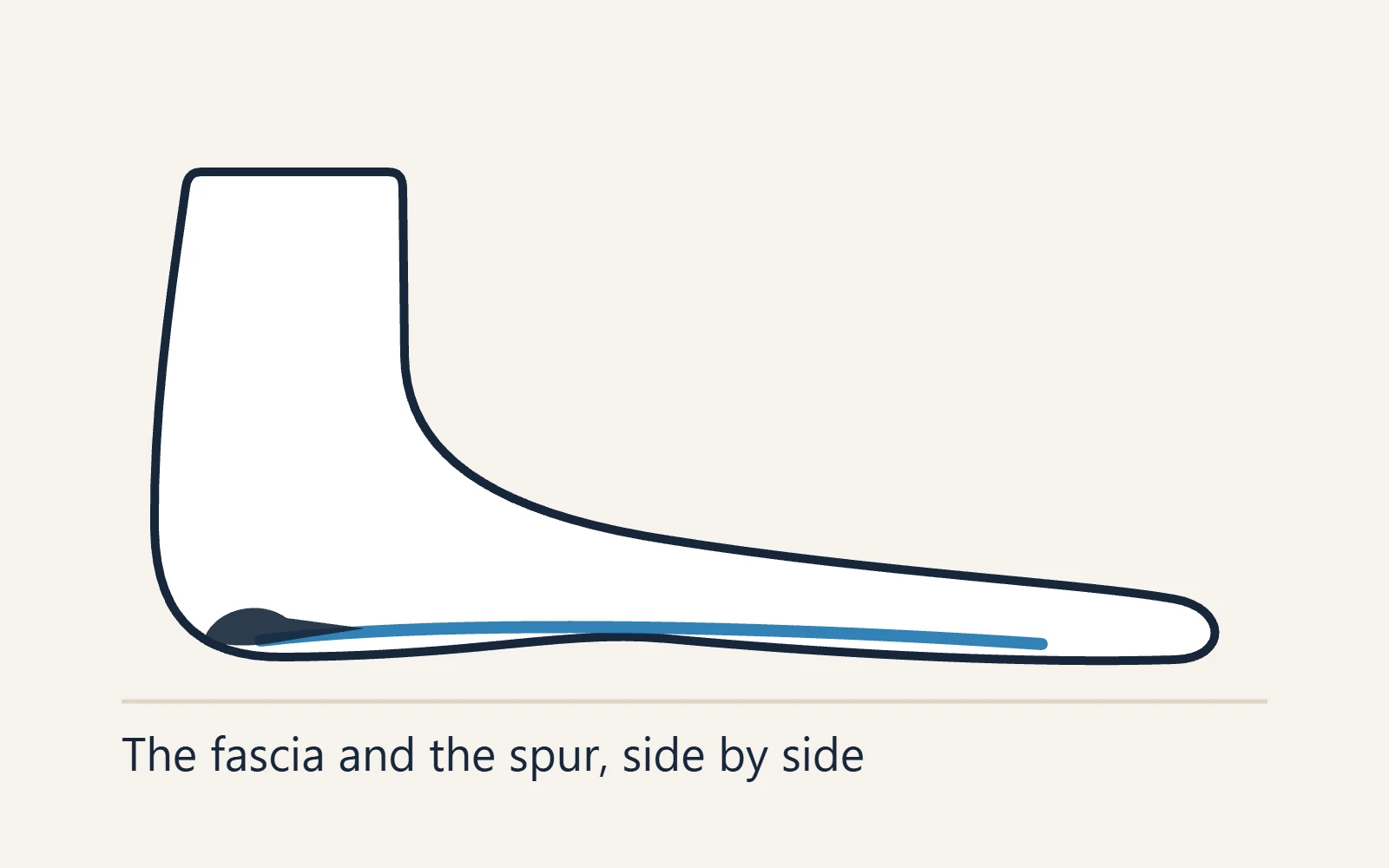
The two things get conflated constantly, including by clinicians, so it is worth separating them cleanly. Plantar fasciitis is a problem of soft tissue at the point where the plantar fascia attaches to the heel bone. A heel spur is a bony outgrowth at roughly the same place. They frequently occur together. That does not make one the cause of the other, and it does not make the spur the source of the pain.
On this page
What each one actually is
The plantar fascia is a thick band running from the heel bone to the base of the toes, working as a tie-rod across the arch. In plantar fasciitis the tissue at its heel attachment becomes degenerative rather than simply inflamed — disorganised collagen, thickening, and micro-tearing at the interface between tendon-like tissue and bone. That interface is called an enthesis.
A heel spur is new bone laid down at that same enthesis in response to years of traction. Bone responds to sustained mechanical load by adding material along the line of that load, which is why spurs point forwards along the direction the fascia pulls. Anatomical studies of spur formation have found that the new bone develops within the enthesis complex, often more closely associated with the origin of the short flexor muscle of the toes than with the plantar fascia itself.
The evidence that the spur is not the pain
- Plenty of people have a spur and no pain whatsoever. Calcaneal spurs are reported in something like one in ten to one in four adults with no heel symptoms at all. If the spur were the pain generator, that would be difficult to explain.
- Plenty of people have plantar fasciitis and no spur. A substantial proportion of symptomatic heels show no spur on X-ray.
- The pain resolves while the spur stays put. The overwhelming majority of plantar fasciitis settles with stretching, loading, footwear and time. The spur is still visible on a repeat X-ray years later, in a heel that no longer hurts.
- Removing it is not necessary. Modern surgery for refractory plantar heel pain addresses the fascia. Where a spur is excised it is usually incidental to the procedure, and it is not what determines the outcome.
- Spur size does not track with pain severity. Large spurs in comfortable heels and small or absent spurs in severely painful ones are both routine.
The useful way to read a spur is as a timestamp rather than a diagnosis. It tells you the fascia has been pulling hard on that attachment for years. It does not tell you what is hurting today, and it does not change the treatment.
Side by side
| Plantar fasciitis | Heel spur | |
|---|---|---|
| What it is | Degenerative change in soft tissue at the fascia’s attachment | New bone laid down at the same attachment over years of traction |
| Where it hurts | Inner front of the underside of the heel, a spot you can cover with a fingertip | Usually nothing — most are silent |
| Morning pattern | Sharp on the first steps, eases over five to twenty minutes | No pattern of its own |
| Shows on X-ray | No — the tissue change is invisible on plain film | Yes, clearly |
| Shows on ultrasound or MRI | Yes — thickening and degenerative change at the attachment | Yes, incidentally |
| Is it the pain source | Yes, in the large majority of plantar heel pain | Rarely, on the underside of the heel |
| Treatment | Load reduction, fascia-specific stretching, progressive loading, footwear and insoles | None required in its own right |
| Does it need removing | Not applicable | Almost never, on the plantar surface |
So why does the report mention it?
Because it is there, and a radiology report describes what is visible. A plain X-ray cannot show fascial degeneration, so on a film of a painful heel the spur is often the only abnormal finding available to name. That is a limitation of the test rather than a diagnosis, and it is the reason so many people leave an appointment believing they have a bone problem when they have a soft tissue one.
X-rays are still worth doing in the right circumstances — to look for a stress fracture, a tumour or an unusual bony cause when the history does not fit. Their role is to rule things out, not to confirm plantar fasciitis.
When a spur genuinely does matter
There are real exceptions, and they are mostly in a different part of the heel.
- Spurs at the back of the heel, where the Achilles attaches, behave differently. Bone there can rub against the tendon and the bursa and genuinely cause symptoms — see Haglund’s deformity and heel bursitis.
- A very prominent plantar spur under a thinned heel pad. If the natural cushioning has atrophied, a bony prominence can become a pressure point. The treatment is still cushioning rather than excision.
- A fracture through a large spur, which is genuinely rare and usually follows a specific injury.
- Spurs as part of an inflammatory arthritis, where enthesitis at multiple sites is the real story and the management belongs to rheumatology.
What to treat instead
If your heel hurts on the underside and is worst on the first steps out of bed, treat it as plantar fasciitis and ignore the spur. That means reducing the daily load, changing what you wear on your feet including indoors, stretching the fascia specifically before standing, and then loading the tissue progressively so it tolerates more. The treatment sequence sets out the order, and the timeline sets expectations.
Have it assessed rather than assumed if any of these apply.
Heel pain that worsens through the day rather than easing after the first few minutes.
Burning, numbness or tingling in the heel or sole.
Pain when the heel is squeezed firmly from side to side.
Both heels, with prolonged morning stiffness, other joints involved, or a history of psoriasis.
Heel pain after a fall from height or in someone with osteoporosis.
The practical upshot
A spur on your X-ray is not good news and not bad news. It is a sign of long-standing traction at the attachment, it is extremely common in people with no symptoms, and it does not need to be removed. The thing worth your attention is the soft tissue, and that responds to consistent mechanical treatment rather than to surgery on a piece of bone that was probably never the problem.
Watch
Dr. Biernacki separates plantar fasciitis, heel spurs and heel stress fractures — and explains which one actually causes pain.
Spur or no spur, you treat the fascia. Dr. Tom’s free 14-day plan from The Heel Pain Playbook starts with a 60-second safety check and a pain map, then five moves with photos and a tracker that shows by day 14 whether it is working. Get the free 14-day plan.
Common questions
Is my heel pain from the spur or the plantar fascia?
Almost certainly the fascia. Heel spurs are common findings in people with no pain at all, plenty of painful heels have no spur, and the pain reliably resolves with the spur still in place. If your pain is on the underside of the heel and worst on the first steps of the day, treat it as plantar fasciitis regardless of what the X-ray shows.
Do heel spurs need to be removed?
On the underside of the heel, almost never. Where surgery is done for refractory plantar heel pain it addresses the fascia, and any spur removal is usually incidental rather than the point of the operation. Spurs at the back of the heel, where the Achilles attaches, are a different matter and can genuinely need surgical attention.
Can a heel spur go away on its own?
No. Bone that has been laid down does not reabsorb, so the spur stays visible on X-ray indefinitely. That is not a problem, because the spur is not what hurts. The pain can resolve completely while the spur remains exactly as it was.
Did the plantar fasciitis cause the spur, or the other way round?
Neither, quite. Both arise from the same underlying situation: years of high traction load at the attachment of the fascia to the heel bone. The soft tissue responds with degenerative change and the bone responds by adding material. They are two consequences of one process rather than cause and effect.
Does the size of the spur matter?
Not in any way that is useful clinically. Large spurs are found in entirely comfortable heels and severe pain occurs in heels with no spur at all. Spur size does not predict pain, does not predict how long recovery takes, and does not change the treatment.
Should I get an X-ray for heel pain?
Plantar fasciitis is diagnosed from the history and examination, not from imaging, and a plain X-ray cannot show the tissue change. X-rays are useful for ruling other things out — a stress fracture, an unusual bony cause, or heel pain that does not fit the usual pattern. If the history is typical and treatment is working, imaging adds little.
- Kumai T, Benjamin M. Heel spur formation and the subcalcaneal enthesis of the plantar fascia. Journal of Rheumatology, 2002 — the anatomical work on where the spur actually forms.
- Johal KS, Milner SA. Plantar fasciitis and the calcaneal spur: fact or fiction? Foot and Ankle Surgery, 2012.
- American Academy of Orthopaedic Surgeons, OrthoInfo. Plantar Fasciitis and Bone Spurs.
- Koc TA, et al. Heel Pain – Plantar Fasciitis: Revision 2023. Clinical Practice Guidelines, Journal of Orthopaedic & Sports Physical Therapy.
- Allam AE, Chang KV. Plantar Heel Pain. StatPearls, NCBI Bookshelf.
- American Academy of Orthopaedic Surgeons, OrthoInfo. Heel Pain.
Related heel pain guides
- Plantar fasciitis
- Heel spurs
- Plantar fasciitis treatment
- Heel pain in the morning
- Haglund’s deformity (bump at the back of the heel)
- Calcaneal stress fracture
- Heel pain: how to work out what you have
- Heel spur treatment
- Heel spur surgery
Heel pain in Michigan?
Dr. Biernacki sees heel pain at Balance Foot & Ankle in Howell and Bloomfield Township. If you have been told a spur is the problem and treatment aimed at it has not helped, an assessment can establish which tissue is actually responsible.
This page is education, not a diagnosis. Read our editorial policy for how it was written and what it will not claim.