Heel pain causes
Insertional Achilles Tendonitis: Pain Where the Tendon Meets the Heel
When the sore spot is on the heel bone itself, the Achilles is being squeezed as well as pulled. That explains why the usual stretches often make it worse, why it is slow, and what settles it.
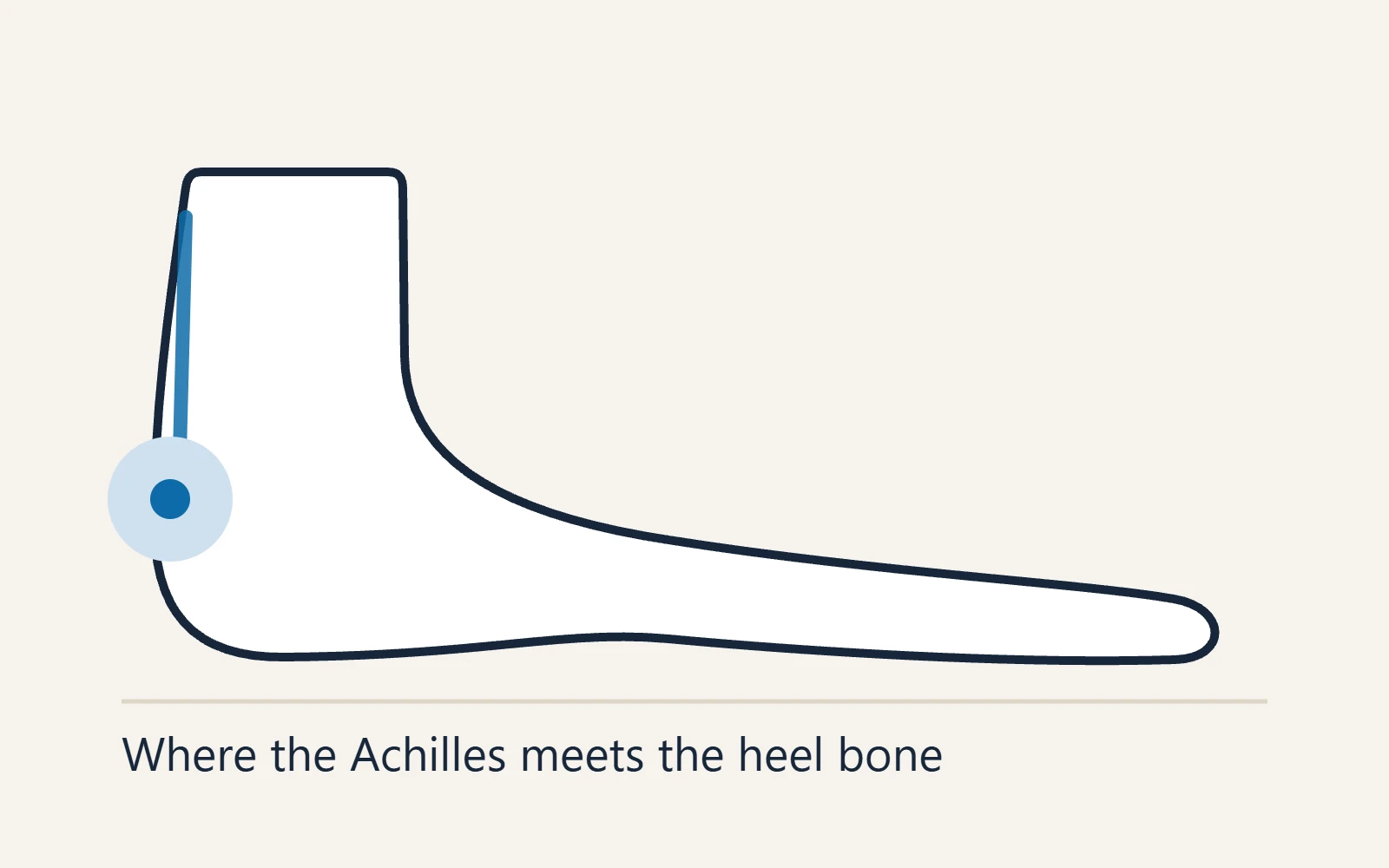
Insertional Achilles tendonitis is pain and degeneration where the Achilles tendon anchors into the back of the heel bone, rather than in the cord above it. Most people get better without surgery by taking the squeeze off the tendon — a small heel lift in both shoes, shoes that do not press on the spot, no deep calf stretching — and strengthening the calf from the floor rather than off a step, over three to six months.
It accounts for roughly a third of Achilles tendon pain and is the more stubborn type. Not sure which type you have? Start with Achilles tendonitis, which explains both.
On this page
Where it hurts, and why
The pain sits on the back of the heel bone, at the attachment or within about two centimetres of it, rather than in the cord above. It is tender to press, stiff after rest and sore with activity, and shoes rub it.
The reason is geometry. The tendon wraps over the upper back corner of the heel bone and attaches about two centimetres lower. Whenever the ankle bends upwards — uphill, on stairs, in a squat, or with the heel dropped off a step — its deep surface is pressed against that corner while the calf pulls along its length. The insertion is squeezed and stretched at once, and a thickened tendon is squeezed harder.
So the classic heel-drop programme off a step, which suits pain higher up the tendon, disappoints here: only 28 to 33% of people with insertional pain rated it good or excellent.
The bump, the bursa and the spur
Three other things share the same few centimetres and often turn up together. All three are also found in heels that do not hurt, so a finding on an X-ray or scan is a clue, not a verdict.
The bursa
A small fluid-filled cushion between the tendon and the bone, just above the attachment. When it is inflamed, squeezing the hollows either side of the tendon hurts. See heel bursitis.
The bump
A Haglund’s deformity is a prominent upper back corner of the heel bone. Heel-bone shape has mostly not differed between people with and without insertional pain, so the bump alone does not explain it. See Haglund’s deformity.
The spur and the calcium
“Achilles heel spur” usually means new bone at the attachment; calcium inside the end of the tendon is calcific insertional tendinopathy. In one study, spurs were almost as common on the pain-free side (55%) as on the painful side (65%), but longer on the painful side.
The first steps of treatment, below, are the same for all three. The spur under the heel is a separate problem (heel spurs).
Who gets it
It turns up at every activity level. Runners are well represented, but many people with it do not run, and in the studies cited on this page the typical patient was middle-aged. A tight calf is a common contributor, and anything that bends the ankle up under load or presses on the back of the heel can aggravate it: more hills, speed work or jumping, flatter shoes, a stiff heel counter. In a growing child, pain here is usually Sever’s disease.
Insertional or something else?
Four problems share the back of the heel, and where your finger lands does most of the sorting.
| Problem | Where it hurts | Tell-tale sign | What helps first |
|---|---|---|---|
| Insertional Achilles tendonitis | On the bone where the tendon attaches, or within about 2 cm of it | Worse uphill, on stairs and when stretching off a step | Heel lift, open-backed shoes, floor-level loading |
| Midportion Achilles tendonitis | In the cord, roughly 2–6 cm above the bone | A thickened segment you can pinch | Graded loading, including heel drops off a step |
| Retrocalcaneal bursitis | The hollows either side of the tendon, just above the bone | Pain when those hollows are squeezed | Heel lift; less upward bend of the ankle under load |
| Haglund’s deformity | A firm bump at the upper back corner of the heel | Hurts in some shoes and not others | Change the back of the shoe |
They often coexist. Stress fractures, ruptures and rarer causes are in back of heel pain.
How it is diagnosed
Mostly by examination: finding the tender spot, squeezing either side for the bursa, looking for a bump, and checking how far the ankle bends up with the knee straight and bent, since a tight calf changes the plan.
A side-on X-ray taken standing shows a spur, calcium and the shape of the heel bone. Ultrasound or MRI shows the tendon and bursa, and earns its place when the result would change the plan: a doubtful diagnosis, or before surgery, when the amount of damaged tendon decides the operation. Scans also find “tendinopathy” in some pain-free tendons, so they are read alongside the examination.
Treatment, in order
The best evidence for this approach is a 2025 randomised trial in 42 sport-active adults. Both groups did a progressive calf programme, but one limited how far the ankle bent, skipped calf stretching and wore heel lifts. By 24 weeks, 18 of 19 in that low-compression group were satisfied, against 11 of 19 who trained into full upward bend and stretched daily.
- Turn the load down, not off. Drop hills, speed work and jumping first; keep walking, and cycle or swim to stay fit. Complete rest calms the pain but leaves the tendon weaker for the return.
- Put a small heel lift in both shoes. It reduces how far the ankle bends with each step, easing the pull and the squeeze; the trial used 12 mm lifts as part of its package. Both shoes, to avoid a leg-length difference.
- Take the pressure off the back of the heel. A rigid heel counter on the sore spot keeps it irritated whatever else you do; wear soft-backed or open-backed shoes for a while.
- Start with holds. If it is irritable, rise partway onto both toes and hold. The trial began with five 30-second holds, building towards a minute.
- Move to floor-level heel raises. Rise on both feet, shift onto the sore leg and lower slowly until the heel touches the floor, never below. In the Swedish pilot study that introduced this version, 18 of 27 patients were satisfied and back to their usual activity at about four months.
- Keep the load rising. Add weight, then single-leg raises, still stopping at floor level. The trial went on from heavier weighted raises to springy hopping exercises, then running and jumping.
- Let pain set the pace. The trial progressed people only when pain stayed below 5 out of 10 during exercise, an hour later and the next morning, and was not rising from week to week. If a rule fails, ease the load for a few days rather than stopping.
For a flare, ice and over-the-counter painkillers, used as directed on the label, can take the edge off; ask a doctor first if you have kidney disease, a stomach ulcer or bleeding, heart failure, take a blood thinner or are pregnant. A short spell in a walking boot can settle a bad flare, but a long one weakens the calf. Night splints are unproven for this type.
The printable programme. Step-by-step instructions for these exercises, with the pain rules and a one-page sheet, are on the Achilles tendonitis exercises page.
What to stop doing
- Heel drops off a step. Letting the heel sink below the step drives the tendon into the bone; the American Academy of Orthopaedic Surgeons advises against them for this type.
- Deep calf stretching, for the same reason; the better group in the trial did none. If you stretch, keep it gentle and stop short of any pinch at the back of the heel.
- A steroid injection into the tendon. It weakens the Achilles and can lead to rupture. One is occasionally placed into an inflamed bursa, deliberately away from the tendon. Platelet-rich plasma and other injections lack good evidence for this type.
Shockwave therapy: what the evidence says
The evidence on shockwave therapy has shifted. A 2008 trial took 50 people whose insertional pain had lasted at least six months: at four months, 64% given shockwave were completely recovered or much improved, against 28% given exercise. But the exercise was the traditional heel drop off a step, and there was no sham treatment.
Sham-controlled trials have not backed it up. In a 2025 trial of 76 people, adding three sessions of radial shockwave to exercise did no better than sham at 6 or 12 weeks, and a 2026 review of nine randomised trials found no clinically meaningful benefit over sham for this type (low to moderate certainty), advised against routine use, and noted two ruptures after the focused kind. If it is offered, treat it as an optional extra alongside loading, never a replacement.
How long it takes
Plan on three to six months, and do not be alarmed by six: this type is harder to settle with exercise than the midportion one. In the 2025 trial most of the gain came in the first 12 weeks. Judge progress by morning stiffness over weeks. If two to three months of consistent work has changed nothing, have the diagnosis and the dose checked; surgery is generally discussed only after six months of proper non-surgical treatment.
Surgery and recovery
A minority need an operation. The usual one removes the degenerate tendon and any calcium, takes off the spur and the Haglund’s prominence, clears the bursa and, where the tendon was detached, reattaches it to the bone with anchors. If more than half the tendon has to be removed, another tendon is moved to the heel for strength, usually the one that bends the big toe (flexor hallucis longus); the big toe still moves afterwards. In a randomised trial of 39 patients over 50, adding that transfer routinely gave more push-off strength but no less pain and no better function.
Recovery is months, not weeks. Most people are allowed to walk in a removable boot or cast within about two weeks, depending on how much tendon was involved. In one surgeon’s series of 166 operations, the average return to activity, including sport, was about seven months, and many people need up to a year of rehabilitation to reach their best. Satisfaction is generally above 87%, but continued pain is the most common complication, in up to 20 to 30%; wound-healing problems, a painful scar and sural nerve injury are the other common ones.
When to be seen
Same day: a sudden pop or snap at the back of the ankle, a feeling of being kicked, sudden weakness pushing off, or being unable to rise onto tiptoe on that leg. Treat it as an Achilles tendon rupture until proven otherwise.
Same day: a hot, red, swollen heel with fever, or broken skin over the heel if you have diabetes or reduced feeling.
Same day: Achilles pain during or after a fluoroquinolone antibiotic, such as ciprofloxacin, levofloxacin or moxifloxacin. The FDA warns that these can damage tendons, the Achilles most often, from within hours of starting to months after finishing, especially over 60, with steroid medicines or after a kidney, heart or lung transplant. Rest the leg and call the prescriber.
This week: pain at rest or at night, any new heel pain if you have diabetes, or both heels in a younger adult with back stiffness that eases with movement (inflammatory heel pain).
More warning signs are on when to worry about heel pain.
Shoes and inserts for this heel. At the insertion, a stiff heel counter pressing on the sore spot can keep it going. Shoes for Achilles tendonitis explains what to look for, and heel lifts reduce compression against the bone.
Common questions
Is insertional Achilles tendonitis the same as a heel spur?
No. A spur at the back of the heel is new bone where the tendon attaches, and it often comes with insertional tendonitis. But spurs are common in heels that do not hurt, the pain comes mainly from the tendon (though a larger spur may add to it), and most people improve without surgery, spur and all. Spurs under the heel: heel spurs.
What is calcific insertional Achilles tendinopathy?
The same condition with calcium deposited in the end of the tendon, seen on a side-on X-ray. The first steps are the same. If surgery is needed, the deposit is removed and the tendon reattached; in the 166-operation series above, people with and without calcification returned to activity in a similar time.
Can I keep running with insertional Achilles tendonitis?
Sometimes: flat, easy and shorter, if the tendon is no worse the next morning, with hills and speed work dropped first. In the 2025 trial, running-based sport waited until the last stage of the programme, at least eight weeks in. See heel pain after running.
How long is recovery after insertional Achilles surgery?
Months. Most people walk in a boot or cast within about two weeks, return to activity averaged about seven months in a 166-operation series, and reaching your best can take up to a year. The more tendon involved, the slower it is.
Related heel pain guides
- Best shoes for Achilles tendonitis
- Achilles tendonitis: the two types
- Achilles tendonitis exercises (printable)
- Haglund’s deformity
- Heel bursitis
- Back of heel pain
- Heel spurs
- KT tape for Achilles tendonitis
- Heel pain after running
- Achilles tendon rupture
- Heel pain quiz
Insertional Achilles pain that is not settling?
If two or three months of a heel lift, kinder shoes and floor-level loading have not moved it, the diagnosis or the dose usually needs checking. Dr. Tom Biernacki sees these at Balance Foot & Ankle.
Howell — 4330 E Grand River Ave, Howell, MI 48843
Bloomfield Township — 43494 Woodward Ave #208, Bloomfield Township, MI 48302
Monday to Thursday, 9:00am–4:30pm; Friday, 9:00am–2:30pm · (810) 206-1402
Further reading
- Chimenti RL, Cychosz CC, Hall MM, Phisitkul P. Current concepts review update: insertional Achilles tendinopathy. Foot & Ankle International. 2017;38(10):1160–1169. doi:10.1177/1071100717723127
- Pringels L, Capelleman R, Van den Abeele A, et al. Effectiveness of reducing tendon compression in the rehabilitation of insertional Achilles tendinopathy: a randomised clinical trial. British Journal of Sports Medicine. 2025;59(9):640–650. doi:10.1136/bjsports-2024-109138
- Jonsson P, Alfredson H, Sunding K, Fahlström M, Cook J. New regimen for eccentric calf-muscle training in patients with chronic insertional Achilles tendinopathy: results of a pilot study. British Journal of Sports Medicine. 2008;42(9):746–749. doi:10.1136/bjsm.2007.039545
- Rompe JD, Furia J, Maffulli N. Eccentric loading compared with shock wave treatment for chronic insertional Achilles tendinopathy: a randomized, controlled trial. Journal of Bone and Joint Surgery, American Volume. 2008;90(1):52–61. doi:10.2106/JBJS.F.01494
- Alsulaimani B, Perraton L, Vallance P, Powers T, Malliaras P. Does shockwave therapy lead to better pain and function than sham over 12 weeks in people with insertional Achilles tendinopathy? A randomised controlled trial. Clinical Rehabilitation. 2025;39(2):174–186. doi:10.1177/02692155241295683
- Korakakis V, Kotsifaki R, Sotiralis Y, Malliaras P. Shockwave therapy for midportion and insertional Achilles tendinopathy: a nail in the coffin? A systematic review with meta-analysis. Journal of Orthopaedic & Sports Physical Therapy. 2026;56(5):282–299. doi:10.2519/jospt.2026.13985
- Saxena A, Maffulli N, Jin A, Isa E, Arthur WP, Wahl A. Insertional Achilles tendinopathy: analysis of 166 procedures and return to activity. Journal of Foot and Ankle Surgery. 2021;60(6):1117–1123. doi:10.1053/j.jfas.2021.01.011
- American Academy of Orthopaedic Surgeons, OrthoInfo. Achilles Tendinitis. Accessed October 2026.
- U.S. Food and Drug Administration. Moxifloxacin prescribing information, boxed warning and section 5.2, Tendinitis and Tendon Rupture. Revised July 2025.
This page is education, not a diagnosis. See our editorial policy, and if the pain began with a pop, get it checked the same day.